Abdominal Gunshot Wound
Gastrointestinal, Trauma
First 5 Minutes
- Before patient arrives, if possible BE PREPARED. Obtain information from EMS on route, find patient ID on your EMR, have help ready including sufficient nursing staff, RT etc. If appropriate contact your surgical team and imaging team ahead of time. Clearly assign roles for your trauma team.
- Prepare necessary equipment including ultrasound for prompt FAST scan, chest tube, etc.
- Ensure team is wearing appropriate PPE.
- Bullet trajectory can involve abdomen, chest, flank and pelvis.
- Prompt type and screen in preparation for possible massive transfusion protocol once patient has arrived. Have 2 units of uncrossmatched blood ready at the bedside.
- EMERGENCY LAPAROTOMY INDICATIONS:
- Peritonitis signs.
- Hemodynamic instability.
- Evisceration.
- Hematemesis, hematochezia.
- If there is no in-house surgical or trauma service, transfer should be completed as soon as patient is stabilized and should NOT be delayed for a full work-up, including imaging and lab work.
Context
- Gunshot wounds (GSWs) are responsible for 90% of abdominal penetrating mortality.
- Hemorrhage is the #1 preventable trauma death cause and may occur rapidly (~2–3 hours after presentation).
- Approximately 30% of exsanguination can occur before hypotension occurs.
- Most likely injuries include small bowel and colon followed by liver. Isolated RUQ liver injuries may be observation candidates.
- GSW are generally NOT locally explored due to difficulty in following wound tracks.
- Damage can differ depending on the type of firearm used.
- Shotgun GSWs may be more superficial depending on the range fired from.
- Rifle or handgun GSWs may have deeper penetration.
- Larger calibre bullets may damage adjacent structures without contact through the shock of impact.
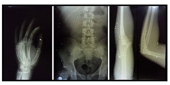
Images gathered from: Search media [Internet]. Wikimedia.org. [cited 2023 Dec 1]. Available from: https://commons.wikimedia.org/w/index.php?search=abdominal+gunshot&title=Special:MediaSearch&go=Go&type=image
Diagnostic Process
- Initiate ATLS algorithm.
- AMPLET Hx simultaneously: Allergies, Meds, PMHx, Last ate; Last menstrual period, Events surrounding injury; Tetanus status.
- History:
- What happened
- Number of gunshots
- Type of firearm used (rifle, handgun, shotgun)
- Proximity of the shooter
- Approximate blood loss at the scene.
- PMHx
- Past psychiatric history if suicide attempt known or suspected
- Medications (specifically blood-thinners)
- Laboratory:
- CBC, blood type and crossmatch, Cr, Glu, ABG if hemodynamic instability, coagulation studies, LFT’s
- Ensure MOVIEE:
- Monitors
- Oxygen administration
- Vital signs
- IV (2 large bore IV’s)
- Exposure
- ECG
Evaluation and Treatment
- Primary survey including POCUS.
- Early massive transfusion protocol activation improves outcomes. Avoid resuscitation with crystalloids in severe hemorrhage, instead prioritizing FFP and PRBCs at a ratio of 1:2 to 1:1 (1), or plasma to platelets to PRBC ratio of 1:1:1 (2) (1:1:1 is the accepted ratio but the local MTP will determine this) Large bore IV access is usually needed including central veins). Avoid patient hypothermia when transfusing – blankets and blood warmer/rapid transfuser.
- If patient is not in extremis, crystalloid resuscitation with an initial bolus of 20mL/kg is appropriate, however this should try to be avoided unless patient is VERY stable.
- In most trauma situations give TXA as a 2g bolus (alternate 1 g over 10 min and 1 G infused over 8 hours). Important to give < 3 hours.
- Fibrinogen supplementation is sometimes done, usually by giving cryoprecipitate by targeting the level of fibrinogen.
- If patient is known to be on blood thinners, reversal should be pursued if possible.
- Provide anesthesia or analgesia as needed while avoiding hemodynamic compromise. Good options include ketamine, midazolam and fentanyl.
- Once stabilized send for whole body CT scan
- If patient remains unstable, transfer to OR for exploratory laparotomy, THEN whole-body CT unless local trauma team dictates otherwise.
- Secondary survey includes full body assessment including the perineum and axilla. All patients with abdominal GSW should have a DRE to assess for hematochezia.
- If patient is not an operative candidate, transfer to ICU.
Emergent operative management if:
- Peritoneal signs.
- Hemodynamic instability.
- Evisceration.
- Hematemesis, hematochezia.
- If operative: Single broad spectrum antibiotic coverage (Cefazolin/Cefoxitin/Ceftriaxone 2 G IV + Clindamycin 600 mg /Metronidazole 500 mg IV) is indicated with both aerobic and anaerobic coverage (3).
Non-operative management if:
- Frequent observation can be performed for at least 24 hours.
- Hemodynamic stability throughout their care.
- No hollow visceral or large vascular injuries.
- Rapid transport to OR is available if condition quickly changes.
- If non-operative: No clear benefit from prophylactic antibiotics described.
Criteria For Hospital Admission
All patients should be admitted to hospital for at least 24 hours of observation.
Criteria For Transfer To Another Facility
- Consult your nearest trauma centre if patient management is beyond the capacity of your medical centre.
- Transfer should be completed as soon as patient is stabilized and should NOT be delayed for a full work-up including imaging and lab work.
Criteria For Close Observation And/or Consult
- Peritonitis signs.
- Hemodynamic instability.
- Evisceration.
- Hematemesis, hematochezia.
Criteria For Safe Discharge Home
After observation period of at least 24 hours, possible discharge:
- Hemodynamic stability.
- No concerning signs of symptoms (peritoneal signs).
- No hematochezia.
- No other major injury.
Quality Of Evidence?

High
We are highly confident that the true effect lies close to that of the estimate of the effect. There is a wide range of studies included in the analyses with no major limitations, there is little variation between studies, and the summary estimate has a narrow confidence interval.
Moderate
We consider that the true effect is likely to be close to the estimate of the effect, but there is a possibility that it is substantially different. There are only a few studies and some have limitations but not major flaws, there are some variations between studies, or the confidence interval of the summary estimate is wide.
Low
When the true effect may be substantially different from the estimate of the effect. The studies have major flaws, there is important variations between studies, of the confidence interval of the summary estimate is very wide.
Justification
Consensus opinion dominates evidence.
Related Information
Reference List
Chang R, Holcomb JB. Optimal fluid therapy for traumatic hemorrhagic shock. Crit Care Clin [Internet]. 2017 [cited 2023 Dec 4];33(1):15–36. Available from: http://dx.doi.org/10.1016/j.ccc.2016.08.007
Cannon JW, Khan MA, Raja AS, Cohen MJ, Como JJ, Cotton BA, et al. Damage control resuscitation in patients with severe traumatic hemorrhage: A practice management guideline from the Eastern Association for the Surgery of Trauma. J Trauma Acute Care Surg [Internet]. 2017 [cited 2023 Dec 4];82(3):605–17. Available from: https://pubmed.ncbi.nlm.nih.gov/28225743/
Brand M, Grieve A. Prophylactic antibiotics for penetrating abdominal trauma. Cochrane Libr [Internet]. 2019 [cited 2023 Dec 4];2019(12). Available from: http://dx.doi.org/10.1002/14651858.cd007370.pub4
Forbes J, Burns B. Abdominal Gunshot Wounds. StatPearls Publishing; 2023.
RESOURCE AUTHOR(S)
DISCLAIMER
The purpose of this document is to provide health care professionals with key facts and recommendations for the diagnosis and treatment of patients in the emergency department. This summary was produced by Emergency Care BC (formerly the BC Emergency Medicine Network) and uses the best available knowledge at the time of publication. However, healthcare professionals should continue to use their own judgment and take into consideration context, resources and other relevant factors. Emergency Care BC is not liable for any damages, claims, liabilities, costs or obligations arising from the use of this document including loss or damages arising from any claims made by a third party. Emergency Care BC also assumes no responsibility or liability for changes made to this document without its consent.
Last Updated Dec 13, 2023
Visit our website at https://emergencycarebc.ca
COMMENTS (0)
Add public comment…
POST COMMENT
We welcome your contribution! If you are a member, log in here. If not, you can still submit a comment but we just need some information.